Introduction
Primary biliary cholangitis (PBC), formerly known as primary biliary cirrhosis, is an autoimmune disease which predominantly affects middle-aged females [1]. The characteristic of PBC is the chronic progressive destruction of small intrahepatic bile ducts, leading to cholestasis, portal inflammation, fibrosis, and biliary cirrhosis [1–3]. The serological hallmark of PBC is the presence of anti-mitochondrial autoantibodies (AMAs) [1]. Although the etiology of PBC is still not fully elucidated, the pathogenesis of PBC is closely associated with the loss of immune tolerance against mitochondrial antigens, resulting in the immune-mediated biliary damage [1, 4]. Previous report showed that the numbers of peripheral T helper (Th) cells were elevated, and the activations of peripheral Th cells correlated with the severity of PBC, indicating the involvement of CD4+ Th cells in the pathogenesis of PBC [5]. Liver-infiltrating terminally differentiated CD8+ T cells revealed elevated cytokine production ability and cytotoxicity in a PBC mouse model, suggesting the key role of hepatic CD8+ T cells in the pathogenesis of PBC [6]. However, the regulation of T cells in PBC patients is still not completely understood.
Interleukin 35 (IL-35) is a newly identified IL-12 cytokine family member and consists of two heterodimeric subunits, Epstein-Barr virus-induced gene 3 (IL-27β chain) and IL-12α chain p35. IL-35 is released by CD4+FoxP3+ regulatory T cells, regulatory B cells, and regulatory CD8+ T cells [7, 8]. IL-35 receptor is also a heterodimer, including IL-12 receptor β2 (IL-12Rβ2) and gp130, which is mainly expressed on immune cells, such as T cells, B cells, and monocytes [9]. Thus, signaling through IL-35/IL-35 receptor pathways always regulates immune cell activity to mediate immunosuppression by inhibition of effector T cell function and enhancement of regulatory cell activity [10]. Our previous studies demonstrated that elevated IL-35 contributed to immune cell dysregulation in various liver diseases, including chronic viral hepatitis [11–13], liver cirrhosis [14], acute-on-chronic liver failure [15], and hepatocellular carcinoma [16–18]. Abnormal expression of IL-35 was also found in several inflammatory autoimmune diseases, leading to the onset and development of these diseases [19, 20]. PBC is an organ-specific autoimmune disease [1]. Li et al. revealed lower plasma IL-35 level in PBC patients, and IL-35 was capable of promoting the inhibitory function of regulatory T cells in PBC patients [21]. However, few studies have focused on the IL-35 regulation of T cells in PBC patients. Accordingly, we examined the correlation between IL-35 and clinical index in PBC patients. Subsequently, peripheral CD4+ and CD8+ T cell activity from PBC patients was functionally analyzed in response to exogenous IL-35 stimulation in vitro.
Materials and methods
Patients and controls
This study complied with all relevant national regulations, institutional policies and was in accordance with the tenets of the Helsinki Declaration (as revised in 2013) and has been approved by the Ethics Committee of the Second Hospital of Jilin University (IRB No. 26/06/17 on 07.06.2017). Written informed consents were obtained from all individuals included in this study. The sample size numbers were calculated by Clinical Research Sample Size Calculator. Fifty-one PBC patients and 28 sex- and age-matched healthy individuals were included between July 2020 and July 2021. The diagnosis of PBC was based on established criteria [22]. All patients were treatment-naive and positive for anti-AMAs. Individuals who were coinfected with hepatitis virus, human immunodeficiency virus, or afflicted with alcoholic cirrhosis, autoimmune hepatitis, primary sclerosing cholangitis, other autoimmune diseases, or cancers were excluded from this study. The clinical characteristics of all studied subjects are listed in Table 1.
| Controls | PBC patients | |
|---|---|---|
| Number | 28 | 51 |
| Sex (male/female) | 7/21 | 8/43 |
| Age, median, IQR (years) | 46 (22, 60) | 38 (25, 51) |
| Alanine aminotransferase, median, IQR (IU/L) | 26 (20, 34) | 62 (48, 112) |
| Aspartate aminotransferase, median, IQR (IU/L) | 20 (14, 29) | 122 (71, 209) |
| Total bile acid, median, IQR (µmol/L) | 7.8 (4.1, 20.1) | 70.0 (58.0, 73.7) |
| Alkaline phosphatase, median, IQR (IU/L) | 54 (41, 67) | 124 (86, 171) |
PBC: Primary biliary cholangitis; IQR: Interquartile range.
Plasma and cell sorting
Ethylene diamine tetraacetic acid anti-coagulant peripheral blood samples were collected from all subjects. Plasma was harvested by centrifugation at 1000×g for 10 min. Peripheral blood mononuclear cells (PBMCs) were isolated by density gradient centrifugation using Ficoll-Hypaque (Sigma-Aldrich, St Louis, MO, USA). CD4+ T cells were purified using Human CD4 MicroBeads (Miltenyi, Bergisch Gladbach, Germany; Catalog# 130-045-101), while CD8+ T cells were sorted using Human CD8+ T cell Isolation Kit (Miltenyi; Catalog# 130-117-027) as previously described [14].
Cell stimulation and culture
The regular bottom cell culture plates were coated with anti-human CD3 epsilon antibody (R&D System, Minneapolis, MN, USA; Catalog# MAB100-500, Clone UCHT1; final concentration: 1 µg/mL) and anti-human CD28 antibody (R&D System; Catalog# MAB342-500, Clone 37407; final concentration: 1 µg/mL) overnight at 4 ∘C as previously described [14]. Sorted CD4+ and CD8+ T cells were then added into coated plates, and cultured with phytohaemagglutinin (PHA) (final concentration: 1 µg/mL) in the presence or absence of recombinant human IL-35 (Peprotech, Rocky Hill, NJ, USA; Catalog# 200–37; final concentration: 1 ng/mL) as previously used [11–18]. The human intrahepatic biliary epithelial cells (HIBECs) (ScienCell, San Diego, CA, USA) were stably transfected with pcDNA3.1-HLA-A*0201 plasmid and were incubated in RPMI 1640 supplemented by 10% fetal bovine serum, 100 U/L penicillin, and 0.1 mg/mL streptomycin in the presence of G418 antibiotic (final concentration: 6.5 mg/mL) at 37 ∘C under 5% CO2 conditions. In certain experiments, CD8+ T cells, which were sorted from 9 HLA-A*02 restricted PBC patients, were stimulated with recombinant human IL-35 for 24 h in the presence of anti-CD3/CD28 antibody. 104 of IL-35 stimulated CD8+ T cells from HLA-A*02 restricted PBC patients were co-cultured in direct contact or indirect contact with 5 × 10⁴ of pcDNA3.1-HLA-A*0201 stably transfected HIBECs in the presence of anti-human CD3/CD28 antibody as previously described [12, 14, 16]. Cells and supernatants were harvested for further experiments 48 h post co-culture.
Enzyme-linked immunosorbent assay
To analyze IL-35 concentration in the plasma and cytokine concentrations in the cultured supernatants, cytokines and cytotoxic molecules levels were measured using commercial enzyme-linked immunosorbent assay (ELISA) kits (CUSABIO, Wuhan, Hubei Province, China), including human IL-35 ELISA kit (Catalog# CSB-E13126h, sensitivity 15.6 pg/mL), human interferon γ (IFN-γ) ELISA kit (Catalog# CSB-E04577h, sensitivity 1.56 pg/mL), human IL-9 ELISA kit (Catalog# CSB-E04642h, sensitivity 3.9 pg/mL), human IL-17 ELISA kit (Catalog# CSB-E12819h, sensitivity 15.6 pg/mL), human IL-22 ELISA kit (Catalog# CSB-E13418h, sensitivity 19.5 pg/mL), human tumor necrosis factor α (TNF-α) ELISA kit (Catalog# CSB-E04740h, sensitivity 1.95 pg/mL), human perforin-1 ELISA kit (Catalog# CSB-E09313h, sensitivity 0.078 ng/mL), human granzyme B ELISA kit (Catalog# CSB-E08718h, sensitivity 0.39 pg/mL), and human granulysin ELISA kit (Catalog# CSB-E09936h).
Flow cytometry
The absolute T cell counts and immune-checkpoint receptors expression on CD8+ T cells were analyzed by flow cytometry, which was performed as previously described [14]. CD3+, CD4+, and CD8+ T cell counts were analyzed using BD Tritest CD4 fluorescein isothiocyanate (FITC)/CD8 phycoerythrin (PE)/CD3 peridinin chlorophyll protein (PerCP) (BD Tritest, San Jose, CA, USA; Catalog# 340298) in a BD TruCount tube (BD Tritest). Harvested CD8+ T cells were stained with allophycocyanin Cy7 mouse anti-human CD8 (BD Pharmingen, San Jose, CA, USA; Catalog# 557834, Clone SK1), FITC mouse anti-human programmed death-1 (PD-1) (BD Pharmingen; Catalog# 557860, Clone MIH4), and PE mouse anti-human cytotoxic T-lymphocyte-associated protein-4 (CTLA-4) (BD Pharmingen; Catalog# 557301, Clone BNI3). Isotype controls were used to enable precise compensation and confirm antibody specificity. Data were acquired using a FACS Calibur flow cytometer (BD Biosciences Immunocytometry Systems, San Jose, CA, USA) and were analyzed using FlowJo software Version 10 (TreeStar, Ashland, OR, USA).
Real-time reverse transcription-polymerase chain reaction
mRNA relative levels corresponding to IL-35 receptor subunits and transcription factors were analyzed by real-time reverse transcription polymerase chain reaction (RT-PCR), which was performed as previously described [11]. Briefly, total RNA was extracted using TRIzol reagent (Invitrogen, Carlsbad, CA, USA; Catalog# 15596-018). First-strand cDNA was synthesized with oligo(dT) primer using PrimeScript RT Master Mix (TaKaRa, Beijing, China; Catalog# RR036B). Real-time PCR was performed using TB Green Premix Ex Taq (TaKaRa; Catalog# RR42WR). The relative gene expressions were quantified using ABI7500 System Sequence Detection Software (Applied Biosystems, Foster, CA, USA). The primers for IL-35 receptor subunits, including IL-12Rβ2 and gp130, were purchased from BioRad (Hercules, CA, USA; Catalog# qHsaCID0006511 and qHsaCID0007540). Other primer sequences were obtained from previously published literature [11, 23].
Cytotoxicity of HIBECs
To analyze the cytotoxicity of HIBECs, lactate dehydrogenase (LDH) level in the supernatants was measured at the end of the incubation period using LDH Cytotoxicity Assay Kit (Beyotime, Wuhan, Hubei Province, China; Catalog# C0016) as previously described [14]. The LDH level in the cultured HIBECs was determined to be “low-level control,” whereas the LDH level in Triton X100-treated HIBECs was determined to be “high-level control.” The percentage of HIBECs death was calculated using the following equation:
(experimental value − low level control)/(high level control − low level control)×100% [14].
Statistical analysis
Data were analyzed using SPSS23.0 for Windows (IBM Corp, Armonk, NY, USA). The Shapiro-Wilk test was used for the normal distribution assay. Data following normal distribution were presented as mean ± standard deviation. Statistical analyses were performed using Student’s t test or paired t test. Pearson correlation analyses were used for correlation analysis for normal distribution data. Data following skewed distribution were presented as median and interquartile range (IQR). Statistical analyses were performed using Mann–Whitney U test or Wilcoxon matched pairs test. Spearman correlation analyses were used for correlation analysis for skewed distribution data. A P value less than 0.05 was considered as statistical difference.
Results
Plasma IL-35 level was reduced in PBC patients
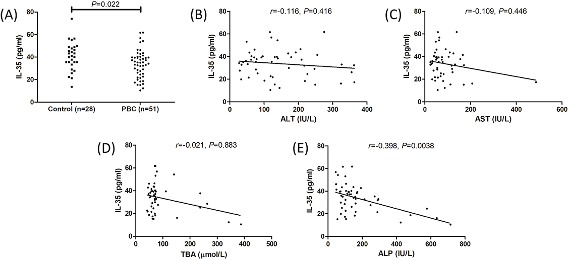
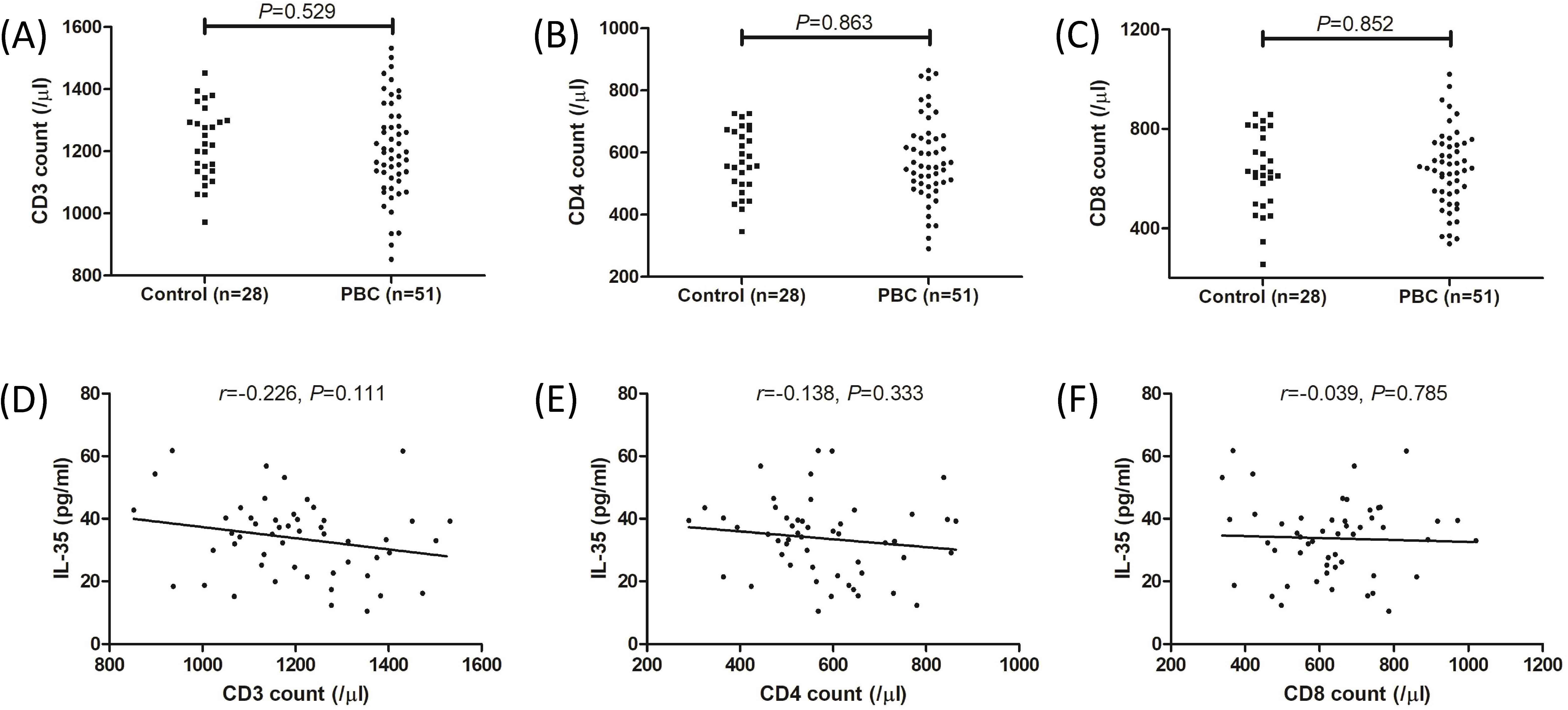
We first examined plasma IL-35 concentration by ELISA. Plasma IL-35 concentration was significantly lower in PBC patients compared to controls (33.72 ± 12.27 pg/mL vs 40.55 ± 12.65 pg/mL; P ═ 0.022, Figure 1A). There were no statistical correlations between plasma IL-35 level and alanine aminotransferase (ALT) (r ═ –0.116, P ═ 0.416, Figure 1B), aspartate aminotransferase (AST) (r ═ –0.109, P ═ 0.446, Figure 1C), or total bile acid (TBA) (r ═ –0.021, P ═ 0.883, Figure 1D). Plasma IL-35 level was negatively correlated with alkaline phosphatase (ALP) (r ═ –0.398, P ═ 0.0038, Figure 1E). Peripheral T cell counts were measured by flow cytometry. There were no remarkable differences in either CD3+ T cell count (1224 ± 118.2/µL vs 1203 ± 155.5/µL; P ═ 0.529, Figure 2A), CD4+ T cell count (572.9 ± 104.2/µL vs 578.0 ± 135.2/µL; P ═ 0.863, Figure 2B), nor CD8+ T cell count (630.0 ± 159.3/µL vs 636.9 ± 155.8/µL; P ═ 0.852, Figure 2C) between controls and PBC patients. Plasma IL-35 level in PBC patients did not correlate with CD3+ T cell count (r ═ –0.226, P ═ 0.111, Figure 2D), CD4+ T cell count (r ═ –0.138, P ═ 0.333, Figure 2E), or CD8+ T cell count (r ═ –0.039, P ═ 0.785, Figure 2F).
Peripheral CD4+ and CD8+ T cells presented stronger inflammatory and cytotoxic phenotype in PBC patients
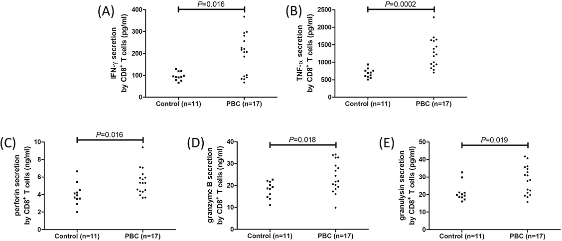
Peripheral CD4+ and CD8+ T cells were sorted from 11 controls and 17 PBC patients and were cultured with anti-CD3/CD28 antibody and PHA for 12 h. Compared with those from controls, CD4+ T cells from PBC patients had elevated mRNA relative levels corresponding to Th1 transcription factor T-bet (2.02 [IQR 1.49, 2.82] vs 1.21 [IQR 1.02, 1.33]; P ═ 0.0006, Figure 3A), Th9 transcription factor PU.1 (4.35 [IQR 2.88, 6.24] vs 1.58 [IQR 1.02, 1.69]; P ═ 0.0001, Figure 3), Th17 transcription factor retinoid-related orphan receptor γt (RORγt) (3.58 [IQR 2.37, 5.40] vs 1.60 [IQR 1.33, 1.71]; P ═ 0.0001, Figure 3C), and Th22 transcription factor aryl hydrocarbon receptor (AhR) (4.00 [IQR 2.09, 6.24] vs 1.42 [IQR 1.33, 1.63]; P< 0.0001, Figure 3D). Similarly, the concentrations of secreting cytokines by CD4+ T cells were higher in the supernatants of cultured CD4+ T cells from PBC patients when compared with controls, including Th1-related cytokine IFN-γ (107.7 ± 14.48 pg/mL vs 82.07 ± 28.76 pg/mL; P ═ 0.0043, Figure 3E), Th9-related cytokine IL-9 (135.4 ± 16.11 pg/mL vs 114.7 ± 14.72 pg/mL; P ═ 0.0021, Figure 3F), Th17-related cytokine IL-17 (64.94 [IQR 57.50, 79.61] pg/mL vs 55.00 [IQR 31.79, 64.36] pg/mL; P ═ 0.025, Figure 3G), and Th22-related cytokine IL-22 (136.7 [IQR 81.32, 269.4] pg/mL vs 68.64 [IQR 42.15, 94.49] pg/mL; P ═ 0.0097, Figure 3H). CD8+ T cells from PBC patients secreted the increased amount of proinflammatory cytokines compared with controls, including IFN-γ (206.8 [IQR 93.09, 268.9] pg/mL vs 93.72 [IQR 78.84, 118.7] pg/mL; P ═ 0.016, Figure 4A) and TNF-α (1264 ± 421.7 pg/mL vs 687.4 ± 128.0 pg/mL; P ═ 0.0002, Figure 4B), as well as cytotoxic molecules, including perforin (5.44 ± 1.49 ng/mL vs 4.05 ± 1.23 ng/mL; P ═ 0.016, Figure 4C), granzyme B (23.98 ± 7.08 pg/mL vs 18.11 ± 3.75 pg/mL; P ═ 0.018, Figure 4D), and granulysin (28.21 ± 8.11 pg/mL vs 21.33 ± 5.17 pg/mL; P ═ 0.019, Figure 4E).
In vitro recombinant human IL-35 stimulation inhibited IL-17 and IL-22 production by CD4+ T cells from PBC patients
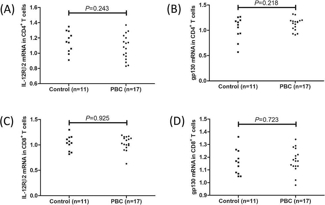
There were no significant differences in IL-35 receptor subunits (IL-12Rβ2 and gp130) in CD4+ T cells between controls and PBC patients (Figure S1A and S1B). Peripheral CD4+ T cells, which were sorted from 12 PBC patients, were cultured with anti-CD3/CD28 antibody and PHA in the presence or absence of recombinant human IL-35 for 12 h. Recombinant human IL-35 stimulation did not affect T-bet and U.1 mRNA relative levels (Figure 5A and 5B) or IFN-γ and L-9 secretion (Figure 5E and 5F) in CD4+ T cells from PBC patients. Exogenous IL-35 stimulation reduced RORγt mRNA relative level (3.28 [IQR 2.09, 5.06] vs 4.36 [IQR 2.49, 6.83]; P ═ 0.034, Figure 5C) and IL-17 production (59.00 [IQR 50.43, 96.23] pg/mL vs 66.58 [IQR 55.85, 104.8] pg/mL; P ═ 0.0098, Figure 5G) in CD4+ T cells from PBC patients. Similarly, IL-35 also notably downregulated IL-22 secretion by CD4+ T cells (99.54 [IQR 70.90, 195.4] pg/mL vs 125.4 [IQR 78.56, 226.8] pg/mL; P ═ 0.0020, Figure 5G). AhR mRNA relative level in CD4+ T cells was also slightly reduced in response to IL-35 stimulation, but this difference failed to achieve statistical significance (4.99 [IQR 2.36, 6.12] vs 5.19 [IQR 2.24, 7.70]; P ═ 0.054, Figure 5D).
In vitro recombinant IL-35 stimulation suppressed the cytotoxicity of CD8+ T cells from PBC patients
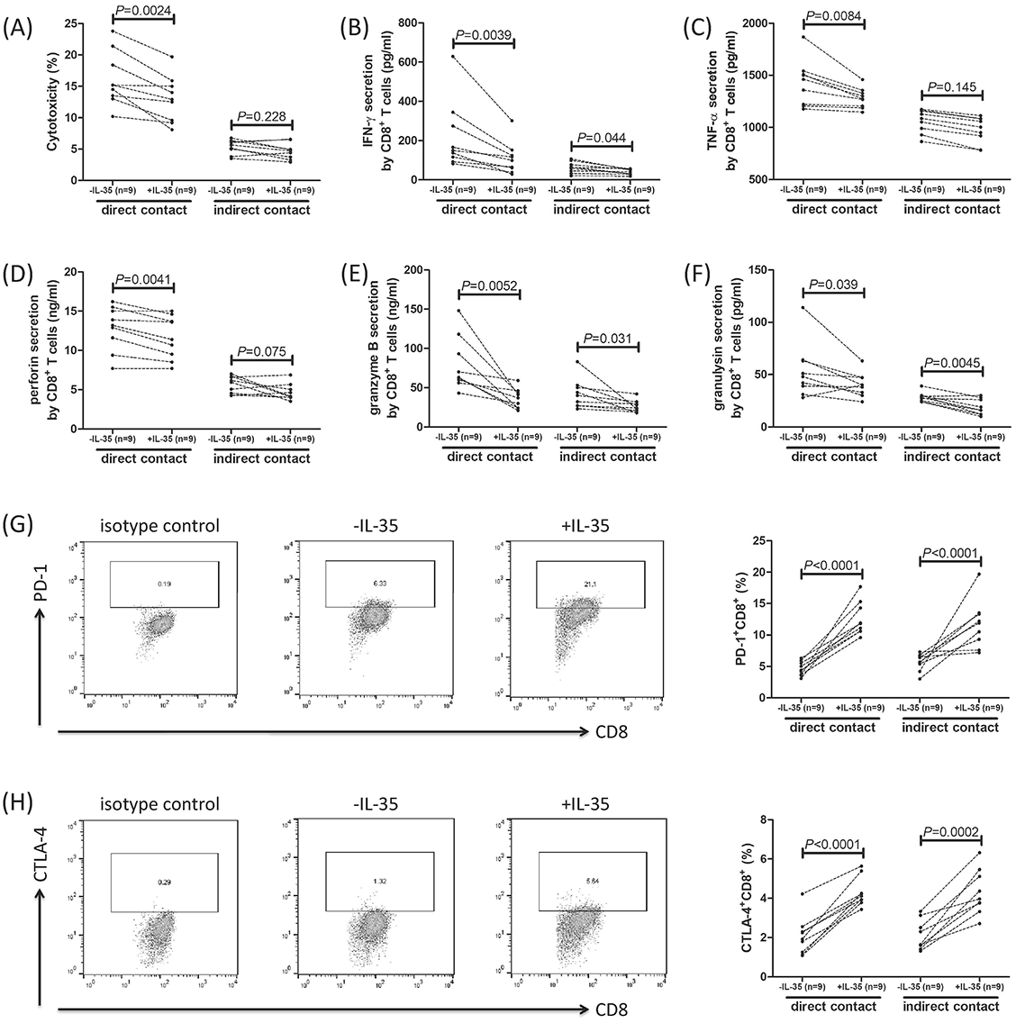
There were no significant differences in IL-35 receptor subunits (IL-12Rβ2 and gp130) in CD8+ T cells between controls and PBC patients (Figure S1C and S1D). In direct contact co-culture system, IL-35-stimulated CD8+ T cells induced lower percentage of HIBECs death when compared with unstimulated CD8+ T cells (12.99 ± 3.68% vs 16.13 ± 4.30%; P ═ 0.0024, Figure 6A). Similarly, IL-35-stimulated CD8+ T cells secreted lower levels of IFN-γ (P ═ 0.0039, Figure 6B), TNF-α (P ═ 0.0084, Figure 6C), perforin (P ═ 0.0041, Figure 6D), granzyme B (P ═ 0.0052, Figure 6E), and granulysin (P ═ 0.039, Figure 6F). However, there was no significant difference in induced HIBECs death between CD8+ T cells with and without IL-35 stimulation in indirect contact co-culture system (4.67 ± 1.28% vs 5.38 ± 1.12%; P ═ 0.228, Figure 6A). IFN-γ (P ═ 0.044, Figure 6B), granzyme B (P ═ 0.031, Figure 6E), and granulysin (P ═ 0.045, Figure 6F) secretion was lower in IL-35-treated CD8+ T cells in indirect contact co-culture system, but IL-35 stimulation did not affect TNF-α (P ═ 0.145, Figure 6C) or perforin (P ═ 0.075, Figure 6D) production in indirect contact co-culture system. The expression of PD-1 and CTLA-4 was determined in CD8+ T cells with and without IL-35 stimulation. Representative flow dots analyses are shown in Figure 6G and 6H. The percentage of CD8+ T cells expressing PD-1 and CTLA-4 was notably increased in response to IL-35 stimulation in both direct contact and indirect contact co-culture systems (P < 0.0001, Figure 6G and 6H).
Discussion
In this study, we observed that circulating IL-35 level was significantly lower in PBC patients, and negatively correlated with ALP. Peripheral CD4+ and CD8+ T cells revealed stronger inflammatory and cytotoxic phenotype in PBC patients, exhibiting increased cytokines secretions and higher cytotoxic molecules productions. Although plasma IL-35 level did not correlate with absolute T cell counts, in vitro exogenous IL-35 stimulation suppressed T cell activity in PBC patients, which presented as inhibiting of IL-17/IL-22 production by CD4+ T cells and dampening cytotoxicity of CD8+ T cells. The current finding indicated an immunosuppressive property of IL-35 in PBC. Insufficient IL-35 expression might be closely associated with enhanced T cell function, leading to immune dysfunction in PBC patients.
Cytokine-mediated immune response plays a crucial role in the pathogenesis of varieties of autoimmune diseases. IL-35 was implicated in the pathogenesis of systemic lupus erythematosus, rheumatoid arthritis (RA), multiple sclerosis, psoriasis, autoimmune hepatitis, etc. [24]. IL-35 level and IL-35-secreting cells were found to be decreased in patients with systemic lupus erythematosus [25, 26], multiple sclerosis [27], and Graves disease [28], revealing the immunosuppressive activity of IL-35 in different types of connective tissue diseases, which was mainly through regulating Th17 cells and regulatory T cells [29]. However, Lian et al. [30] showed that IL-35 subunits were elevated in liver tissue and were positively associated with degrees of hepatic inflammation and fibrosis in patients with autoimmune hepatitis. Furthermore, IL-35 induced expansion of myeloid-derived suppressor cells in liver microenvironment, resulting in a potential protective activity of IL-35 in autoimmune hepatitis [30]. Similarly, serum level of IL-35 was higher in psoriatic arthritis patients, which was characterized by bone erosion in peripheral joints and bone formation in spinal joints [31]. IL-35 was also upregulated at the sites of inflammation in RA synovium, which was even higher than in psoriatic arthritis synovium [32]. Moreover, IL-35 stimulated osteoblasts differentiation activated by TNF-α [33]. In turn, TNF-α induced the elevation of IL-35 secretion by PBMCs, which further promoted the release of IL-1β and IL-6 in RA patients [32], suggesting the proinflammatory potential of IL-35 in RA pathogenesis. Thus, the opposing mechanisms of anti-inflammatory and proinflammatory function of IL-35 might be related to the ultimate development of autoimmune disorder, and in a context dependent manner [20]. Our current results were consistent with the previous finding, revealing the downregulation of circulating IL-35 in PBC patients [21]. Importantly, lower plasma IL-35 was accompanied by higher ALP level in PBC patients. ALP, the representative marker for cholestasis, was localized in both canalicular and basolateral membranes and in the entire cytoplasm of bile duct epithelial cells, and was strongly increased in liver tissue compared with controls and patients with chronic hepatitis [34]. The increased expression of serum ALP might be the result of the elevation of hepatic ALP in PBC [34]. Thus, the lower level of IL-35 might serve as a marker for cholestasis in PBC. However, the role of IL-35 reduction in PBC patients still needs to be further elucidated.
CD4+ Th cells are involved in the pathogenesis of PBC. Th1 cytokines and chemokines, including IFN-γ, chemokine (C-X-C motif) ligand 9, and IFN-γ-induced protein-10, were increased in PBC patients and were reduced after ursodeoxycholic acid treatment [35]. Decreased liver-infiltrating CD4+ Th1 cells indicated a good response to ursodeoxycholic acid therapy [36]. Th9-secreting cytokine IL-9 was found in higher concentrations in peripheral blood and liver tissues of PBC patients [37]. Th17 population was decreased in the circulation and was associated with greater accumulation in the liver during PBC progression [38]. Deletion of IL-22, which was the hallmark cytokine for Th22 cells, reduced biliary injury in a PBC mouse model [39]. Herein, we found that transcription factors and cytokine productions by CD4+ Th cells were robustly increased in the peripheral blood of PBC patients, indicating the involvement of Th1, Th9, Th17, and Th22 cells in PBC. Peripheral CD4+ Th cells exhibited a stronger inflammatory phenotype in PBC, which might be important for the induction of biliary damage in PBC patients. IL-35 dampened anti-tumor activity of Th1 cells in peripheral blood and bronchoalveolar lavage fluid in patients with non-small cell lung cancer [40]. IL-35 also inhibited Th17 response in various diseases progression, including allergic rhinitis [41], visceral leishmaniasis [42], acute hepatitis B [43], etc. Our previous study also showed that IL-35 suppressed Th9 cell activity in patients with hepatocellular carcinoma [18]. However, the regulatory function of IL-35 to peripheral CD4+ Th cells in PBC was not fully consistent with the role in other diseases. IL-35 level did not show significant correlation with absolute count for CD4+ T cells. In vitro IL-35 stimulation did not affect Th1 and Th9 differentiation, but it suppressed Th17 and Th22 function in PBC patients. This finding indicated that Th17 and Th22 cells might be more sensitive to exogenous IL-35 stimulation in PBC patients and might be a key mediator for the pathogenesis of PBC. Taken together, IL-35 appeared to be an essential regulator for effector CD4+ T cell dysfunction in PBC.
CD8+ T cells mediated cytotoxicity and induced the destruction of biliary epithelial cells [44, 45]. CD8+ T cells revealed cytotoxicity through two independent mechanisms. On the one hand, secretion of cytotolytic molecules played vital roles in CD8+ T cell-induced target cell death, and this process required direct cell-to-cell contact. On the other hand, secretion of proinflammatory cytokines also contributed to cytotoxicity, which was in a cell-to-cell contact independent manner [14, 16]. Circulating CD8+ T cells from PBC patients expressed higher levels of perforin, granzyme B, and granulysin as well as elevated IFN-γ and TNF-α. This finding indicated that peripheral CD8+ T cells exhibited enhanced proinflammatory and cytotoxic phenotype during PBC. Importantly, our previous data suggested that IL-35 induced CD8+ T cell exhaustion in chronic viral hepatitis [12], liver cirrhosis [14], acute-on-chronic liver failure [15], and hepatocellular carcinoma [16]. Our current results revealed that there was no significant correlation between circulating IL-35 expression and CD8+ T cell absolute count. Importantly, in vitro IL-35 stimulation only reduced the CD8+ T cell-mediated target cell death in direct contact co-culture system, but not in the indirect contact co-culture system. This process was accompanied by downregulation of cytotoxic molecules and proinflammatory cytokines, as well as upregulation of immune checkpoint receptors. This finding indicated that cytotoxic molecules, but not proinflammatory cytokines, might be the predominant mediators for CD8+ T cells in PBC. Cytokine-induced cytotoxic might not be sufficient for cytolytic activity to HIBECs. Collectively, IL-35 stimulation suppressed non-specific CD8+ T cell cytotoxicity in PBC.
There were several limitations in the current study. First, we only analyzed peripheral blood but not liver-infiltrating T cell function in PBC. The phenotype of T cells might be altered in liver microenvironment in PBC patients. Second, the in vivo experiments using animal models should be performed to confirm the effect of IL-35 on T cell activity in PBC. Third, we only analyzed the regulatory function of IL-35 on T cells. Myeloid cells and other immune cells also play important roles in mediating PBC. The role of IL-35 on modulating these cells will be further elucidated.
Conclusion
Plasma IL-35 level was reduced, whereas peripheral T cell activity was enhanced in PBC patients. Exogenous IL-35 stimulation in vitro suppressed Th17/Th22 response and inhibited cytotoxicity of CD8+ T cells from PBC patients. Reduced circulating IL-35 might be insufficient to suppress T cell function, leading to the immune dysregulation in PBC patients. Thus, IL-35 may be considered as a therapeutic target for PBC.


